VII.10.A. Atherosclerosis
It has been proven that insulin resistance not only involves a disturbance of the metabolism in the tissues (liver, muscle and fat), but also has effects on the cellular level (platelets, endothelium, macrophages and vascular smooth muscle cells). MetS patients may have cardiovascular complications. These patients are characterized by higher VLDL and LDL and lower HDL levels.
Atherosclerosis is a disease in which the earlier flexible vascular walls harden. Fat is deposited in the vascular walls (atheroma) with age, and increasingly extends and obstructs the blood flow. The first formation of deposits involves lipoprotein molecules adhering to the inner vascular wall where it is damaged (Fig. 27). As a result, the vascular wall becomes more vulnerable, leading to its thickening. Soft plaque is first formed.
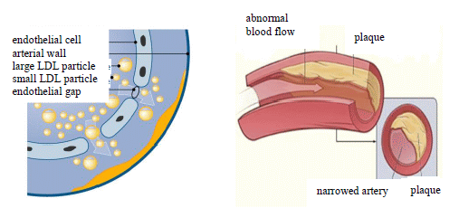
Figure 27. The effect of LDL on the vascular wall, and the development of atherosclerosis
(Fredrickson et al. NEJM 1967; 276: 148)
The structure of the vascular wall is damaged because of the changed endothelial function; it becomes permeable for some molecules and cells (vascular remodeling). The next step is leucocyte adhesion, which leads to the increased production of cytokines and growth factors. An inflammatory reaction develops; thereafter, the deposited fats are formed into plaques. Macrophages release TNF-α and IL-1β. The molecular processes between the macrophages and the endothelium lead to increased affinity of the vascular wall for oxidized LDL.
Monocytes are bound to the vascular wall by the endothelium-produced monocyte chemotactic protein (MCP), and they then turn into macrophages in the subintimal area. Their role is to eliminate the oxidized LDL fat molecules. During the process, the cells are transformed into foam cells filled with lipid. The accumulation of foam cells causes a yellow striped discoloration in the intima (fatty streak). The atheroma plaques contain lipids, cholesterol, cholesterol esters, collagen and muscle items. The deposits of molecules narrow the vascular lumen, and hence obstruct the blood flow.
The released PDGF (platelet-derived growth factor) induces an increase in the amount of vascular wall muscle, and these synthesize collagen and stabilize plaques (fibrous plaque).
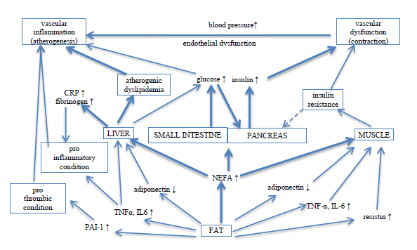
Figure 28 Plaque formation
(Grundy Nature Reviews Drug Discovery 5, 295–306 (April 2006) | doi:10.1038/nrd2005
NEFA = non-esterified fatty acids)
The later-developed hard plaque expands to the three layers of the vessel wall, and the vascular lumen is almost completely blocked.
Damage to the atherosclerotic plaque may lead to the accumulation of thrombocytes on the vascular surface (thrombocyte aggregation), and the decreased vascular lumen may be totally closed by a thrombus (atherothrombosis). Alternatively, the lagging thrombus causes an embolism. The process is the most dangerous in the coronaries (Fig. 28).
Atherothrombosis may lead to sudden cardiac death, infarct or stroke. The regression of advanced plaques is a very long process.