III.5.A. Neurological control of eating
The liver and the adipose tissue provide the brain with information about the energy storage status, the nutrition access in the blood and the GIT, the appearance, taste and odor of the nutrition; the external environmental impacts (such as the availability of food, and social eating habits) and other internal signals (such as mood). The brain integrates the information and the general outputs coordinate the ingestion (search for food, starting a meal and stopping a meal) and the modulation processes (energy storage and release).
Figure 13. Regulation of eating
(PFC = prefrontal cortex, OFC = orbitofrontal cortex; (A)CC = (anterior) cingulate cortex; VTA = ventral tegmental area; NAc = nucleus accumbens; NTS = nucleus tractus solitarius; CCK = cholecystokinin; GLP-1 = glucagon-like peptide-1; PYY = peptide tyrosine tyrosine, insula = part of cerebral cortex.
The Metabolic Syndrome (Kindle Locations 5337-5339). Wiley, Kindle Edition.)
The brain synchronizes two main eating aspects: the “maintenance of homeostasis” and the “award”. The maintenance of homeostasis means body weight maintenance, the filling of the energy stores and the maintenance of proper ion homeostasis; the award means the experience and eating as an award (Fig. 13).
The food uptake is regulated by the hypothalamic, brainstem nuclei and the limbic system.
The main signal adapter center is the hypothalamus (Fig. 14).
the ventromedial hypothalamus – “satiety center” (HT-VMH), and
the lateral hypothalamus – “fasting center” (HT-LH).
The brainstem nuclei (nucleus tractus solitarii) together with other nuclei and tracts control the food intake. Chemosensitive receptors of the brainstem sense the chemical composition of the blood. From among the upper brain areas the limbic system is very important. Other brain regions regulate the hypothalamic and brainstem centers through the sensation of the food taste, odor and quality.
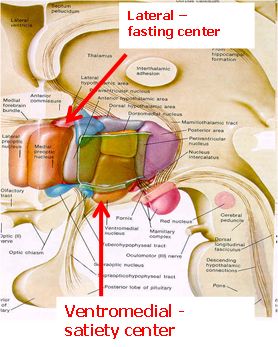
Figure 14. Eating regulatory center in the hypothalamus
During the stimulation of the fasting center (HT-LH), animals seek to feed. Lesion of this area may have the result that the animal does not feed or drink. (The thirst center is injured by such as lesion.) HT-LH is in a relationship with the exercise center.
The cells in the satiety (HT-VMH) center sense the amount of Glu.
During the stimulation of this area, animals do not feed. A lesion of this area can lead to 3-4-fold weight gain. The lesioned animals feed more often, the release of insulin is enhanced, and the fat storage increases.
After the lesioning, two phases occur:
The dynamic phase (4-14 weeks) is characterized by a rapid weight gain (hyperphagia). The physical activity decreases. In the subsequent static phase, the food intake decreases, but the animal remains overweight. Such experimental animals prefer sweet food.
Figure 15. The main centers of eating regulation in the brain
(AGRP = Agouti-related peptide, ARC = nucleus arcuatus, CART = cocaine- and amphetamine-regulated transcript, CCK = cholecystokinin, CRH = corticotropin-releasing hormone, GIT = gastrointestinal tract, GLP-1 = glucagon-like peptide-1, HT-LH = lateral hypothalamus, MCH = melanin-concentrating hormone, NPY = neuropeptide Y, NTS = nucleus tractus solitarius; OX = oxytocin, OXA = orexin, PFA = perifornical area, POMC = pro-opiomelanocortin, PYY = peptide tyrosine tyrosine, PVN = nucleus paraventricularis, TRH = TSH-releasing hormone;
red arrow = inhibition, green arrow = stimulation
Elena Valassi et al, Neuroendocrine control of food intake, Nutrition, Metabolism and Cardiovascular Diseases (2008), 18, 158-168.)
The nucleus arcuatus (ARC) plays an important role in the regulation. The hormones NPY and AGRP, which increase food intake are released from this area. The ARC also contains neurons that produce the hormones POMC and CART, which inhibit food intake. The nucleus paraventricularis (PVN) releases inhibitory hormones (TRH, OX and CRH). The lateral hypothalamus (HT-LH) and the perifornical area (PFA) release food intake-stimulatory hormones (OXA and MCH). Signals from the adipose tissue reach the ARC and activate it to inhibit food intake. This stimulates the other pathway, which increases the food intake (Fig. 15).